Eye Library / Diagnostic Tests / Fundus Fluorescein Angiography
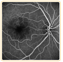
Fundus Fluorescein Angiography: is useful for evaluating various disease effects on the retina. The test requires an injectable dye (fluorescein), specialized camera with filters, and timing. The dye is injected into the patient's arm; within seconds, the dye travels to the blood vessels inside the eye. Photographs are taken to document filling of vessels and any fluid leakage as the dye circulates through the eye. A digital camera is often used which allows the physician to interpret the results immediately. Test does not involve the use of X–rays or harmful forms of radiation. Fluorescein dye is best for studying the retinal circulation. Certain eye disorders, such as diabetic retinopathy and retinal vascular occlusive disease affect primarily the retinal circulation and are usually imaged with fluorescein dye. In other disorders, such as age–related macular degeneration, where leakage is from the deeper choroidal vessels, test is partial useful. Some people may experience slight nausea after dye injection that usually passes quickly. Patients who are allergic to the dye can develop itching and a skin rash. There is also a possibility of an infiltrate of the dye into the skin at the injection site; this would cause some discomfort or discoloring of the skin for several days. Fluorescein dye will also turn a patient’s urine orange and may slightly discolor the skin as well for a brief period.


